")
")
")
Modern microscopes for biological imaging are often "black boxes" whose exact operating principle remains unknown and whose optical resolution and price seem to be inversely proportional to each other. With UC2 (You. See. Too.), a low-cost, 3D-printed, open-source, modular microscopy kit is presented here and its versatility demonstrated by realizing a complete microscope development cycle from the concept to the experimental phase.
Introduction
The brightfield microscope enclosed in an incubator monitors the differentiation of monocyte to macrophage cells at cellular resolution level (e.g. 2 μm) for seven days. In addition, the geometry is transferred by incorporating very few additional components into a 400-euro light sheet fluorescence microscope for volumetric observations of a transgenic zebrafish expressing green fluorescent protein (GFP). The aim is to establish an open standard in optics to facilitate coupling with various complementary platforms. By making the content and comprehensive documentation publicly available, the systems presented here are suitable for simple and uncomplicated replications, modifications and extensions.
Educational: light sheet microscope for education
In this section, the versatility is demonstrated by converting the BF system of the previous chapter into a light sheet microscope (Fig. 2g) by replacing the LED array with a laser pointer, adding a second microscope objective, a beam expander, a cylindrical lens and the specimen stage using a larger base plate. A video explaining the conversion together with a detailed conversion recipe and a detailed schematic of the open SPIM-inspired [15] setup can be found in Supplementary Video 4 and Supplementary Notes 7.7 , respectively. A 3D data stack of zebrafish larvae expressing GFP in the blood vessels was acquired, which was further drift-corrected and deconvolved using the program "GenericDeconvolution" by Heintzmann et al. (available on request) (Fig. 3l, Supplementary Fig. 3) . At the moment the results only show a proof of concept that it is possible to build a light sheet system at such a low price (400 Euro). Better performance would require better optical components and better adaptation to the applications. However, the light sheet microscope has proven its usefulness in teaching and has given users a valuable insight into a method they often work with but only know as a black box. The minimum number of printed and commercially available components required to build the aforementioned setups as well as telescopes, projectors, Abbe diffraction experiments or holographic (e.g. lensless) imaging devices in a cost- and resource-efficient way was analyzed to compile a ready-to-print collection of parts and documentation called "TheBOX" (see supplementary notes 7.11) and a version optimized for microscopy teaching courses called "CourseBOX". It is supported by continuously improved documentation with step-by-step instructions and tutorials. The system was tested at various conferences, workshops and teaching environments (see supplementary notes 9) and a lot of constructive feedback was given to further improve the system. During these iterations, a lower barrier to use and understanding for new workshop participants was observed, which can be attributed to the improvement of the documentation and the steadily increasing robustness of the cubes.
 Beginnend mit einer biologischen Fragestellung/Idee, für die ein bildgebendes Gerät benötigt wird, das in (b) entworfen (invertiertes Inkubatormikros-kop) und in (c) mit UC2-Komponenten aus der CAD-Bibliothek umgesetzt wird. Nach dem Druck und Zusammenbau (d) wird das Gerät in seine Arbeitsumgebung (z.B. Inkubator) gestellt (e) und ist bereit, Langzeit-Bildserien von z.B. MDCK-Zellen aufzunehmen, die in (i) und im ergänzenden Video 6 visualisiert werden. Die Fernsteuerung erfolgt über „smarte Komponenten“ (z.B. Mobiltelefon, Raspberry Pi) in (h). Die Wiederverwendung von Komponenten erlaubt den Umbau in ein Handy-Mikroskop (f) oder Lichtblattmikroskop (g) innerhalb von Minuten (siehe Ergänzungsvideo 5) und Ergänzende Hinweise 7.8). CL: Zylinderlinse, TL: Tubuslinse, L: Laser, LA: LED-Array, M: Spiegel, MO: Mikroskopobjektiv, P-CAM: Detektor (Smartphone oder Raspberry Pi), S: Probenpositioniertisch, F: Emissionsfilter, Z: Fokussiertisch") Fig. 2: Rapid prototyping with UC2. Typical workflow for creating a UC2 application: (a) Starting with a biological question/idea that requires an imaging device, which is designed in (b) (inverted incubator microscope) and realized in (c) with UC2 components from the CAD library. After printing and assembly (d), the device is placed in its working environment (e.g. incubator) (e) and is ready to acquire long-term image series of e.g. MDCK cells, which are visualized in (i) and in the supplementary video 6. Remote control takes place via "smart components" (e.g. cell phone, Raspberry Pi) in (h). Reuse of components allows conversion into a cell phone microscope (f) or light sheet microscope (g) within minutes (see Supplementary Video 5) and Supplementary Notes 7.8). CL: Cylindrical lens, TL: Tube lens, L: Laser, LA: LED array, M: Mirror, MO: Microscope objective, P-CAM: Detector (smartphone or Raspberry Pi), S: Sample positioning stage, F: Emission filter, Z: Focusing stage
Fig. 2: Rapid prototyping with UC2. Typical workflow for creating a UC2 application: (a) Starting with a biological question/idea that requires an imaging device, which is designed in (b) (inverted incubator microscope) and realized in (c) with UC2 components from the CAD library. After printing and assembly (d), the device is placed in its working environment (e.g. incubator) (e) and is ready to acquire long-term image series of e.g. MDCK cells, which are visualized in (i) and in the supplementary video 6. Remote control takes place via "smart components" (e.g. cell phone, Raspberry Pi) in (h). Reuse of components allows conversion into a cell phone microscope (f) or light sheet microscope (g) within minutes (see Supplementary Video 5) and Supplementary Notes 7.8). CL: Cylindrical lens, TL: Tube lens, L: Laser, LA: LED array, M: Mirror, MO: Microscope objective, P-CAM: Detector (smartphone or Raspberry Pi), S: Sample positioning stage, F: Emission filter, Z: Focusing stage
Multimodal: fluorescence and label-free imaging
Although fluorescence imaging was shown to be possible with the UC2 incubator and light sheet configuration (e.g. fluorescence overlay in Fig. 3c), the sensitivity of the Raspberry Pi camera suffered from the high noise contribution, quantified in Supplementary Notes 7.5 , and reduced sensitivity due to the Bayer pattern. Replacing the Raspberry Pi RGB camera with a cell phone with a backlit monochromatic camera (P20 Pro, Huawei, China), which captures up to 4 × more photons, significantly improved imaging performance. A quantitative comparison was performed by imaging mCLING-ATTO 647N (SYSY, Germany) labeled with E. coli using a UC2 laser-based infinite optics fluorescence microscope (×100, NA = 1.25 oil, λexc = 635/637 nm, see Supplementary Notes 7.4 equipped with a Raspberry Pi camera or a cell phone camera was obtained with a standard research microscope (Zeiss Axiovert TV, ×100, NA = 1.46) in Fig. 3i-k . The cell phone camera was able to clearly resolve the plasma membrane of the bacteria (see Fig. 3i-k, small sub-ROI). The practical resolution dcellphone = 0.6 μm was determined in comparison to dRaspi = 1.13 μm and dZeiss = 0.27 μm, under similar experimental conditions (e.g. exposure time, gain, laser intensity) using Fourier ring correlation (FRC) [38] (further quantified in Supplementary Notes 7.4). Using the GUI on the Raspberry Pi, a time-lapse series of moving fixed but mobile (e.g. in aqueous suspension) E. coli bacteria at 1 fps could also be planned using the aforementioned infinity-correlated setup (see Supplementary Video 7).
UC2 also enables the design of more sophisticated systems. As an example, the fabrication of an image scanning microscope (ISM) [39], where the excitation laser in the previous infinity-corrected setup was replaced by a customized module incorporating a laser scanning video projector (Sony MP.CL1A, Japan; Supplementary Notes 7.9). Compare images of GFP-labeled human pulmonary microvascular endothelial cells (HPMEC) acquired with the UC2-ISM (Optika, × 20, NA = 0.4, N-Plan, additional information Supplementary Notes 7.9 ) with a state-of-the-art laser scanning confocal microscope (Leica TCS SP5, Fluotar × 20, NA = 0.5, Germany) in Fig. 3e-h. The computationally reconstructed "superconfocal" image [40] Fig. 3g shows an optical section compared to the wide-field equivalent Fig. 3f.
-b), sind nach 42 h deutlich verlängerte Zellen zu sehen (roter Pfeil). Das Wachstum einer differenzierenden Zelle ist als durchschnittliche Fläche der Zellen über mehrere Zeitschritte und verschiedene Experimente in d) dargestellt. c) Der Hellfeldkanal überlagert mit einem Fluoreszenzsignal von fixierten, mit CellTracker green markierten Makrophagen, aufgenommen mit dem Mikroskop im Inkubator. e) Weitfeld-Fluoreszenz und f) das berechnete „superkonfokale“ Ergebnis von GFP-markierten HPMECs, beleuchtet mit einem Laser-Scanning-Projektor, aufgenommen mit einer Handykamera. Die herangezoomten Bilder zeigen die Verbesserung des optischen Schnitts bei strukturierter Beleuchtung in h) im Vergleich zum Weitwinkel in g), bei dem kleinere Zellstrukturen verloren gehen. i) Ein Vergleich der gleichen Probe, aufgenommen mit einem kommerziellen Laser-Scanning-Konfokalmikroskop. j) Ein aufgenommener z-Stapel einer GFP-exprimierenden Drosophila-Larve. k) Die Verwendung eines LED-Rings als Beleuchtung ermöglicht die quantitative Phasenabbildung von Wangenzellen mit A-IDT. l) LED-Matrizen können schnell zwischen Hell- und Dunkelfeldabbildung umschalten, wie in l) gezeigt") Fig. 3: Visualization of the different imaging modalities from a UC2 setup Variation of macrophage morphology In a)-b), clearly elongated cells can be seen after 42 h (red arrow). The growth of a differentiating cell is shown as the average area of the cells over several time steps and different experiments in d). c) The brightfield channel overlaid with a fluorescence signal from fixed macrophages labeled with CellTracker green, recorded with the microscope in the incubator. e) Widefield fluorescence and f) the calculated "superconfocal" result of GFP-labeled HPMECs, illuminated with a laser scanning projector, recorded with a cell phone camera. The zoomed-in images show the improvement of the optical section with structured illumination in h) compared to the wide angle in g), where smaller cell structures are lost. i) A comparison of the same sample imaged with a commercial laser scanning confocal microscope. j) An imaged z-stack of a GFP-expressing Drosophila larva. k) Using an LED ring as illumination enables quantitative phase imaging of cheek cells with A-IDT. l) LED matrices can quickly switch between light and dark field imaging as shown in l)
Fig. 3: Visualization of the different imaging modalities from a UC2 setup Variation of macrophage morphology In a)-b), clearly elongated cells can be seen after 42 h (red arrow). The growth of a differentiating cell is shown as the average area of the cells over several time steps and different experiments in d). c) The brightfield channel overlaid with a fluorescence signal from fixed macrophages labeled with CellTracker green, recorded with the microscope in the incubator. e) Widefield fluorescence and f) the calculated "superconfocal" result of GFP-labeled HPMECs, illuminated with a laser scanning projector, recorded with a cell phone camera. The zoomed-in images show the improvement of the optical section with structured illumination in h) compared to the wide angle in g), where smaller cell structures are lost. i) A comparison of the same sample imaged with a commercial laser scanning confocal microscope. j) An imaged z-stack of a GFP-expressing Drosophila larva. k) Using an LED ring as illumination enables quantitative phase imaging of cheek cells with A-IDT. l) LED matrices can quickly switch between light and dark field imaging as shown in l)
Furthermore, when using an LED matrix (Adafruit #1487, NY, USA) as a light source in transmission mode, the selection of the illumination wavelength, specific patterns for contrast maximization [41] using the openKöhler module (Supplementary Notes 7.10), dark-field illumination (Fig. 3n) or quantitative phase methods such as "(quantitative) differential phase contrast" (qDPC26, see Supplementary Notes 7.6) and "Fourier ptychography microscopy" (FPM [42]). One replaced the matrix with an LED ring (Adafruit#1463) to demonstrate computational refocusing of a recovered phase map of cheek cells (Fig. 3m) to apply "annular intensity diffraction tomography" (aIDT [43], see also Supplementary Notes 7.6 and the reconstructed Z-stack in Supplementary Video 3).
Discussion
In this paper, a modular microscope kit was presented that has the potential to serve as a truly open standard. This standard is defined by the dimensions and shape of the base cube based on a variety of parameters and experience in order to be as general as possible. The goal of not only creating new parts, but defining a common interface for the ever-growing variety of different components has been achieved. By also connecting UC2 to existing rail and cage systems from Thorlabs, Newport, Edmund Scientific and the like, as well as to existing lab equipment, it makes it easier for users to connect and reuse existing components and setups, reinforcing the idea as an open standard.
The inherent versatility of the UC2 toolbox was demonstrated by first realizing an entire life cycle of a microscope in a few steps for an incubator-enclosed brightfield configuration and then presenting examples of how different modern microscopic techniques can be implemented by replacing a few components.
But of course, there are also limits to the long-term stability of 3D-printed setups due to the temperature-dependent deformability of PLA and ABS materials. The iterative design process resulted in an interchangeable mechanical module with minimal bending, which can be actively supported by an autofocus routine or manual refocusing. This allowed long-term stability to be achieved in several experiments where imaging was performed with 4 incubator-enclosed microscopes over 7 days without significant focus drift, while in vitro macrophage differentiation was continuously monitored. The possibility of long-term measurements allowed us to replicate the data published by Xia et al [37], in which the elongated shape of macrophages is correlated with their movement. The incubator-integrated microscope proved the advantages of its inherently small footprint and high throughput capability by parallelizing experiments on a very low budget while providing tailored imaging tools for e.g. microfluidic chips or within highly secure biological environments (BSL3+).
Another important limiting factor in fluorescence imaging (e.g. light-sheet setup) is the performance of the used Raspberry Pi camera (v2.1), which can be improved with more sensitive camera sensors, e.g. from cell phones or industrial cameras. Therefore, rather than being a productive imaging tool, the light-sheet system is a low-cost (≈400 Euro) proof-of-concept approach that provides a valuable insight into the method for educational users.
With "TheBox", a sophisticated toolset for teaching purposes has been introduced. Together with a set of ready-to-use documentation, optical concepts (interference, image formation, etc.) and a variety of light microscopy methods, it provided an open-access microscopy platform at a price between 100 and 600 Euros. This gives students and end users the opportunity to learn how modern optical methods work and encourages interdisciplinary approaches where several teaching topics are covered simultaneously. Exemplary teaching material is shown in the supplementary figure 3 .
The UC2 toolbox can be easily integrated into existing frameworks such as Openflexure stage [16], Micro-manager [44] and ImJoy [42] due to its inherent modularity on both the hardware and software side. In addition, the existing pool of ready-to-use modules enables rapid prototyping in optics, teaching and other areas. A versatile, flexible, expandable enabling tool is urgently needed in optics. With UC2, scientists hope to create an optical equivalent of what Arduino is to electronics and Fiji [46] is to biomedical imaging by making cutting-edge microscopy techniques accessible to everyone. Efforts have been made to counteract the difficulties of reproduction by providing step-by-step protocols at the hardware level to directly reproduce experiments. It is believed that the community addressed will take up the UC2 toolbox as a true open standard, supporting the easier dissemination of laboratory research and rapid system prototyping not only in research but also in education.
Methods Production of components and selection of additional parts for the incubator-integrated microscope
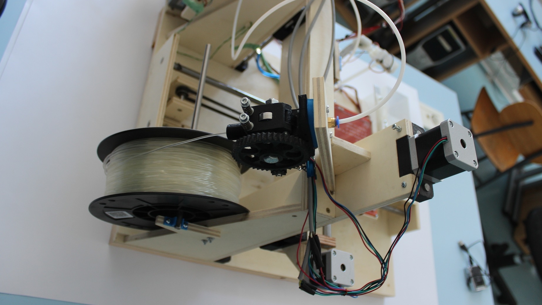
Readers can find a detailed description of each individual part as well as the bill of materials (BOM) in the Supplementary Notes 1, available in the GitHub repository at http://github.com/bionanoimaging/UC2-GIT. In general, all components of the UC2 toolbox are designed with common CAD software (Autodesk Inventor 2019, MA, USA; OpenSCAD 2019.05) and printed with commercially available FDM-based 3D printers (Prusa i3, MK3s, Czech Republic; Ultimaker 2+/3, Netherlands), using PLA (Tprint = 215 °C) as the printing filament in all cases except for the Z-stage and the base plate. The infill was chosen between 20 -40 %, together with a layer height of 0.15 mm, which provided sufficient precision and stability for fully optical build-ups. The monolithically printed Z-stage cube (Supplementary Notes 7.1) based on a linear or flexurally mounted and horizontally mounted base plate for use in the incubator was printed with ABS, which gave better long-term stability at Tincubator = 37 °. The Z-stage adapts to common objective lenses (i.e. RMS thread), which are linearly shifted using a worm drive realized with an M3 screw and nut and driven by a low-cost stepper motor (28BYJ-48, China).
The black material was used in most cases to reduce stray light or unwanted reflection and scattering. To decontaminate the printed parts, the assembled cubes were sprayed with 70% ethanol before being transferred to the Live Cell Imaging Laboratory (LSB2, UKJ Jena) facility.
For the magnetic locking mechanism, 5 mm neodymium sphere magnets were pressed into the printed base plate, which adapted to galvanized M3 × 12 mm cap screws (Würth M3 × 12, ISO 4762/DIN 912) located in each side of the cube to ensure a stable connection. Additional wires added to the magnets and screws, respectively, carry electro-optical modules (e.g. LED array) with electrical voltage (i.e. 5V, GND), while a rectifier prevented issues with wrong polarity.
To keep the optical design simple and compact, an inexpensive (15 euros) infinity-corrected objective lens (×10, NA = 0.3, China) was used, with the light beam folded using a cosmetic mirror (20 cents). The image obtained at a reduced tube length (dtube = 100 mm) was captured using a back-illuminated CMOS sensor (Raspberry Pi Camera, v2.1, UK) connected to a Raspberry Pi v3B. An additional module containing a pair of motorized, low-cost XY microtables (3 Euro, dx = dy = ±1.2 mm, Aliexpress, China) for precise positioning of the sample in XY (Supplementary Notes 7.1)
can be used. For brightfield and quantitative imaging, an 8 × 8 LED array (Adafruit #1487, NY, USA) was used, running a GUI on a 7-inch touchscreen (Raspberry Pi, UK) that allowed LEDs to be activated individually to maximize contrast according to Siedentopf's principle [41]. For fluorescence imaging of GFP-labeled HPMEC cells, we equipped the fluorescence module (Supplementary Notes 7.1) with two high-power LEDs in dark-field configuration (Cree, 450 nm/405 nm ± 20 nm) and added a gel color filter in front of the CMOS sensor (ROSCO #11). Detailed information about the UC2-ISM can be found in the supplementary notes 7.9 .
Hardware synchronization and image acquisition
All sources together with the full documentation of the software, briefly described below, as well as detailed instructions can be found in our GitHub repository and in Supplementary Notes 6.1. A reduction in the number of wires for "active" modules (e.g. equipped with motors, LEDs) was achieved by a microcontroller connected to a wired I2C BUS (Arduino Nano, Italy) or a wireless network based on the MQTT protocol (ESP32 WROOM, China). The Rasp-berry Pi v3B was chosen as the master device for the 4-wire I2C connection. The ESP32 can be controlled with any MQTT device, e.g. Raspberry Pi, cell phone or other ESP32/Arduino microcontrollers on the same network, allowing remote control of the device (e.g. from the office).
A user-friendly Python-based [47] GUI running on a 7-inch touchscreen allows access to functions such as scheduling experiments, setting up imaging modalities (e.g. illumination patterns) and hardware/frame synchronization for different applications (e.g. microscope in an incubator). Images from the camera module (Raspberry Pi, v2.1) are saved as compressed JPEG images to save disk space or to have the unprocessed RAW Bayer pattern data written to EXIF metadata. In cases where cell phones (e.g. P9/P20 Pro, Huawei, China) were used as imaging devices, the open-source camera APP FreeDCam [48] was used to have full control over the imaging parameters (i.e. ISO, exposure time) and access to the RAW images. USB batteries (power storage) enabled autonomous operation in rural areas for several days.
Live drift correction was tested to account for the expansion of the material through software-based autofocus (i.e. axial defocusing). A direct spatial filter (i.e., Tennengrad) [49] was used as the focusing metric and a variance-based filter was used as the image sharpness metric (Supplementary Notes 7.3).
Image analysis and image processing
A customized Python script [47] processes long-term measurements (e.g. one frame per minute over 1 week) by storing the RAW data in binning and creating a preview video. Then a reference frame, in which the lateral sample shift appears to have settled, and ROI (regions of interest) for fixed image features - here: Dirt on the sensor - are defined. In a second iteration, image statistics such as min, max, mean or sharpness and shift are calculated using cross-correlation estimation for the whole image and the ROIs with respect to the reference frame. ABS, which has a large linear thermal expansion coefficient of 70 × 10-6/K50, tends to deform particularly dominantly during the one-hour heating phase in the incubator. Dark and damaged images were excluded using the statistical measures. To compensate for XY drift, shifts were applied and an image stack average was calculated for the green channel. Only the green channel was further processed. Flat-fielding and dirt correction were achieved by dividing by the mean of the entire stack after background correction to account for uneven illumination and sensor errors (e.g. dirt, scratches). Fiji (v1.53c46) was used to measure cell size (i.e. macrophages, see Supplementary Fig. 2), diameter was determined manually over 10 time frames across the entire 1-week measurement of all four microscopes. In each frame, a single cell was manually selected before the roundness factor was calculated with a customized macro.
For task-specific image processing directly on the cell phone, such as processing the ISM measurements or frame segmentation, the cloud-based image processing framework ImJoy (v0.11.1545), available on the GitHub repository (Supplementary Note 6.1) , was used.
For the quantitative phase measurements based on the aIDT, the publicly available Matlab (2017b, The MathWorks, MA, USA) code by Li et al [43] was used with minor modifications according to the optical system using the cell phone microscope (see Supplementary Note 7.6) .
Possible fluctuations of Z-stacks imaged with the light sheet microscope were registered with a cross-correlation based routine before deconvolution based on the publicly available program "GenericDeconvolution" by Heintzmann et al. (available on request) removed the blurring.
Sample preparation
Peripheral blood mononuclear cells (PBMCs) were isolated from blood donated by healthy volunteer adult donors by Ficoll density centrifugation. The study and the experimental protocols used were approved by the Ethics Committee of the University Hospital Jena (assigned study number 2018-1052-BO). Briefly, blood was mixed with isobuffer (phosphate buffered saline, PBS without Ca/Mg (Gibco, Darmstadt, Germany), 2 mM ethylenediaminetetraacetic acid (EDTA, Sigma Aldrich, Steinheim, Germany), 0.1% bovine serum albumin (BSA, Sigma Aldrich, Steinheim, Germany)) and applied to Biocoll (Biochrom, Merck, Germany) without mixing in a 50 ml tube. Biocoll and blood were centrifuged at 800 × g for 20 minutes without breaks. PBMCs were transferred to a new 50 ml tube and washed twice with isobuffer. PBMCs were seeded at a density of 1×10⁶ cells/cm2 in X-Vivo 15 medium (Lonza, Cologne, Germany) supplemented with 10% (v/v) autologous human serum, 10 ng/ml granulocyte-macrophage colony-stimulating factor (GM-CSF) and 10 ng/ml macrophage colony-stimulating factor (M-CSF) (PeproTech, Hamburg, Germany) and Pen/Strep (Sigma Aldrich, Steinheim, Germany). After 1 h, PBMCs were washed twice with Gibco RPMI 1640 medium (Thermofisher, MA, USA), and the remaining monocytes were then rinsed with X-Vivo with supplements. 16 h after isolation, monocytes were washed with pre-warmed (PBS, without Ca/Mg) and incubated for 7 min with pre-warmed with 4 mg/ml lidocaine (Sigma Aldrich, Steinheim, Germany) and 1 mM EDTA. Separated monocytes were placed in a 15-ml tube and centrifuged at 350 × g for 7 min. Sediment monocytes were counted and 1.5 × 10⁵ were seeded into a 35-mm dish and rinsed with 3 ml of X-Vivo 15 with supplements .
Acknowledgments
This study was supported by the Center for Sepsis Control and Care (Federal Ministry of Education and Research (BMBF), Germany, FKZ 01EO1502) and the Leibniz ScienceCampus InfectoOptik Jena, which is funded by the Leibniz Association's Strategic Networking funding line. In addition, this work was financially supported by the German Research Foundation via the Cluster of Excellence "Balance of the Microverse" as part of the Excellence Strategy-EXC 2051-Project-ID 690 390713860 and by the European Commission via Marie Skłodowska-Curie Actions (MSCA) Innovative Training Network EUROoC (Grant No. 812954) to A.S.M. The authors would like to thank the Lichtwerkstatt Jena-Open Photonics Makerspace at Friedrich Schiller University Jena for sharing resources and facilities for several workshops. Also to Leibniz IPHT Jena e.V. for funding the project through the Innovation Fund. Human pulmonary microvascular endothelial cells transfected with eGFP (HPMEC-eGFP), kindly provided by Dr. Lothar Koch and Andrea Deiwick from the Institute of Quantum Optics at Leibniz University. The authors would like to thank Nora Mosig, Melanie Ulrich and Tobias Vogt for their excellent technical support. Also Kaspar Podgorski for organizing and HHMI Janelia for funding the UC2 workshop at the HHMI Janelia Research Farms. Thanks are also given to Xian Hu (Edna), Kay Schink, Felix Margadant and Oddmund Bakke for organizing, funding, hosting and preparing Drosophila and MDCK samples for the workshop at the University of Oslo. Dr. Uta Naumann from the Leibniz Institute on Ageing - Fritz Lipmann Institute (FLI) Jena is thanked for providing the zebrafish samples. In addition, Philipp Kahn for creating the UC2 project website, Eda Bingöl for her support during the filming and witelo Jena e.V. for organizing several UC2 workshops. The authors would like to thank Ronny Förster, Tomáš Čižmár, Nico Schramma and Kyriacos Leptos for fruitful discussions. Further thanks go to Øystein Helle from the Arctic University of Tromsø for the preparation and Patrick Then for his help with the imaging of the E. coli bacteria. R.H. gratefully acknowledges the support of the Collaborative Research Center SFB 1278 (PolyTarget, project C04) funded by the German Research Foundation.
Contributions:
B.D., R.L. and S.C. conceived the UC2 idea, B.D., R.L., S.C., B.M., R.H. and H.W. performed data curation, B.D., R.L. and S.C. contributed to formal analysis, B.D., B.M. and H.W. developed hardware components, B.D., R.L. and X.U. developed the acquisition software, B.D., R.L., A.M. and R.H. organized fundraising, B.D., R.H. and R.L. supervised, conceived and planned the project, designed the instrument, interpreted the data and wrote the manuscript. All authors read and approved the final manuscript. See "Authors" for abbreviated names.
This is a translation of the original article in two parts in Nature Communications volume 11, Article number: 5979 (2020), published under creative commons license CC-BY 4.0 http://creativecommons.org/licenses/by/4.0/
Supplementary notes and videos can be found at https://www.nature.com/articles/s41467-020-19447-9
THE AUTHORS
Benedict Diederich (B.D.) 1, 2, 3 ,
René Lachmann (R. L.) 1, Swen Carlstedt (S. C.) 4,
Barbora Marsikova (B. M.) 1, 3 Haoran Wang (H. W.) 1,
Xavier Uwurukundo (X. U.) 1
Alexander S. Mosig (A. M.) 4,
Rainer Heintzmann (R. H.) 1, 2, 3
1 Leibniz Institute of Photonic Technology, Albert-Einstein-Strasse 9, 07745, Jena, Germany
2 Institute of Physical Chemistry and Abbe Center of Photonics, Helmholtzweg 4, Friedrich-Schiller-University, Jena, Germany
3 Faculty of Physics and Astronomy, Friedrich-Schiller-University, Jena, Germany
4 Jena University Hospital, Institute of Biochemistry II, Am Klinikum 1, Jena, Germany
Literature
[15] Pitrone, P.G. et al: OpenSPIM: An open-access light-sheet microscopy platform, Nat. Methods 10, 2014, 598-599
[16] Sharkey, J.P.; Foo, D.C.W.; Kabla, A.; Baumberg, J.J.; Bowman, R.W.: A one-piece 3D printed flexure translation stage for open-source microscopy, Rev. Sci. Instrum. 87, 2016, https://doi.org/10.1063/1.4941068
[17] Maia Chagas, A.; Prieto-Godino, L.L.; Arrenberg, A.B.; Baden, T.: The €100 lab: A 3D-printable open-source platform for fluorescence microscopy, optogenetics, and accurate temperature control during behavior of zebrafish, Drosophila, and Caenorhabditis elegans, PLoS Biol. 15, 2017, e2002702
[18] Cybulski, J.S.; Clements, J.; Prakash, M.: Foldscope: Origami-Based Paper Microscope, PLoS ONE 9, June 2014, e98781
[19] Martens, K.J.A. et al: Visualization of dCas9 target search in vivo using an open-microscopy framework, Nature Communications 10, 2019, 3552
[20] Diederich, B.; Then, P.; Jügler, A.; Förster, R.; Heintzmann, R.: cellSTORMCosteffective super-resolution on a cellphone using dSTORM. PLoS ONE 14, 2019, e0209827
[21] Winters, B.J.; Shepler, D.: 3D printable optomechanical cage system with enclosure, HardwareX 3, 2018, 62-81
[22] Delmans, M.; Haseloff, J.µ.: Cube: A Framework for 3D Printable Optomechanics, J. Open Hardware 2, 2018, 1-9
[23] Arduino, I. Arduino: Open source products for electronic projects, http:// www.arduino.org/, 2019
[24] Inc, R. Raspberry Pi Teach, Learn, and Make with Raspberry Pi, https://www.raspberrypi.org/, 2016
[25] Booth, M.J.: Adaptive optical microscopy: the ongoing quest for a perfect image, Light: Science & Applications 3, 2014, e165-e165
[26] Tian, L.; Waller, L.: Quantitative differential phase contrast imaging in an LED array microscope, Opt. Express 23, 2015, 11394
[27] Gross, H.; Singer, W.; Totzeck, M.; Gross, H.: Handbook of Optical Systems 1-690, https://doi.org/10.1002/3527606688, 2006
[28] Semiconductors, N. UM10204 I 2 C-bus specification and user manual Rev. 64 April 2014 User manual Documen is a machine-to-machine (M2M) Internet of Things http://www.nxp.com
[29] For the Advancement of Structured Information, S. O. MQTT https://mqtt.org/, 2019
[30] Mantovani, A.; Sozzani, S.; Locati, M.; Allavena, P.; Sica, A.: Macrophage polarization: tumor-associated macrophages as a paradigm for polarized M2 mononuclear phagocytes, Trends Immunol. 23, 2002, 549-555
[31] Guilliams, M.; Scott, C.L.: Does niche competition determine the origin of tissue-resident macrophages? Nature Reviews Immunology, https://doi.org/10.1038/nri.2017.42, 2017
[32] Andreesen, R.; Picht, J.; Löhr, G.W.: Primary cultures of human blood-born macrophages grown on hydrophobic teflon membranes, J. Immunol. Methods 56, 1983, 295-304
[33] Jay, S.M.; Skokos, E.; Laiwalla, F.; Krady, M.M.; Kyriakides, T.R.: Foreign body giant cell formation is preceded by lamellipodia formation and can be attenuated by inhibition of Rac1 activation, Am. J. Pathol. 171, 2007, 632-640
[34] Waldo, S.W. et al: Heterogeneity of human macrophages in culture and in atherosclerotic plaques, Am. J. Pathol. 172, 2008, 1112-1126
[35] McWhorter, F.Y.; Wang, T.; Nguyen, P.; Chung, T.; Liu, W.F.: Modulation of macrophage phenotype by cell shape, Proc. Natl Acad. Sci. USA 110, 2013, 17253-17258
[36] Rosania, G.R.; Swanson, J.A.: Microtubules can modulate pseudopod activity from a distance inside macrophages, Cell Motil. Cytoskeleton. 34, 1996, 230-245
[37] Xia, Z.; Triffitt, J.T.: A review on macrophage responses to biomaterials, Biomed. Mater. 1, https://doi.org/10.1088/1748-6041/1/1/R0, 2006
[38] Banterle, N.; Bui, K.H.; Lemke, E.A.; Beck, M.: Fourier ring correlation as a resolution criterion for super-resolution microscopy, J. Struct. Biol. 183, 2013, 363367
[39] Müller, C.B.; Enderlein, J.: Image Scanning Microscopy, Phys. Rev. Lett. 104, 2010, 198101
[40] Heintzmann, R.; Benedetti, P.A.: High-resolution image reconstruction in fluorescence microscopy with patterned excitation, Appl. Opt. 45, 2006, 5037-5045
[41] Diederich, B.; Wartmann, R.; Schadwinkel, H.; Heintzmann, R.: Using machine-learning to optimize phase contrast in a low-cost cellphone microscope, PLoS ONE 13, 2018, e0192937
[42] Ou, X.; Horstmeyer, R.; Zheng, G.; Yang, C.: High numerical aperture Fourier ptychography: principle, implementation and characterization, Opt. Express 23, 2015, 5473-5480
[43] Li, J. et al: High-speed in vitro intensity diffraction tomography, Advanced Photonics 1, 2019, 1-13
[44] Edelstein, A.; Amodaj, N.; Hoover, K., Vale, R.; Stuurman, N.: Computer Control of Microscopes Using µ Manager, Curr. Protoc. Mol. Biol. 92, 2010, 14.20.1-14.20.17
[45] Ouyang, W.; Mueller, F.; Hjelmare, M.; Lundberg, E.; Zimmer, C.: ImJoy: an open-source computational platform for the deep learning era, Nat. Methods 16, 2019, 1199-1200
[46] Schindelin, J. et al: Fiji: An open-source platform for biological-image analysis, Nat. Methods https://doi.org/10.1038/nmeth.2019, 2012
[47] Harris, C.R. et al: Array programming with NumPy. Nature 585, 2020, 357-362
[48] Fuchs, I.: Github: FreedCam https://github.com/KillerInk/FreeDcam, 2019
[40] Royer, L.A. et al: Adaptive light-sheet microscopy for long-term, high-resolution imaging in living organisms, Nat. Biotechnol. 34, 2016, 1267-1278
[49] Inc, O. Omnexus Plastics and Elastomers https://omnexus.specialchem.com/polymerproperties/properties/coefficient-of-linear-thermal-expansion, 2020
