Few developments accelerate the gain in knowledge as much as those in the field of imaging. Current developments in computed tomography, X-ray imaging and detection and data analysis are described below.
Since the development of photography, new imaging technologies have always led to progress in the understanding of the processes under investigation. In medical imaging, imaging technology, computer-aided image analysis and adaptive therapy are closely linked.
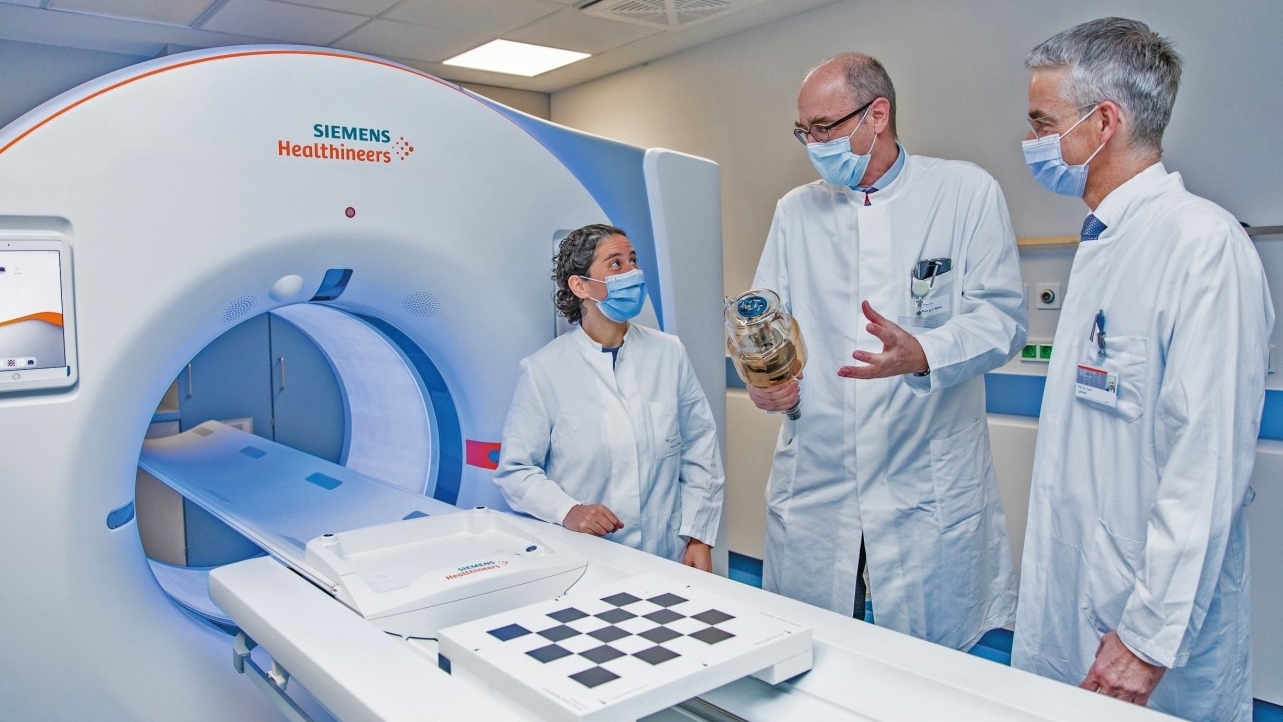
Quantum counting computer tomograph [1]
The computer tomograph (CT) is the workhorse of radiologists. This is because most radiological examinations with cross-sectional images, for example after accidents or for the diagnosis of diseases, are carried out using CT devices. There are two main goals in the further development of this technology: Better images and a lower radiation dose for patients.
The new Naetom Alpha quantum counting CT scanner is a huge step forward in this respect. The first 20 units worldwide are currently being put into operation - one of them at the Institute of Diagnostic and Interventional Radiology at Hannover Medical School (MHH).
Around 12 million CT scans are performed in Germany every year. Conventional CT detectors first convert X-rays into visible light, which is then transformed into an electric current. This energy is then used to generate a digital image. However, important information is lost during the intermediate step of converting light into electricity. This results in reduced image contrasts and reduced image sharpness. The new quantum counting computer tomograph from Siemens Healthineers works with a fundamentally different technology. It converts the X-ray photons, i.e. the light quanta, directly into electrical signals. The intermediate step is no longer necessary. The new X-ray detector is able to count the individual light quanta in each pixel. Hence the name "quantum-counting computer tomograph". The direct transformation into electrical current means that the energy information is retained. The images are sharper and richer in contrast and provide new and revealing information. A much more differentiated impression can be gained and it is possible to see exactly whether contrast media, soft tissue or bones are being imaged, for example.
The images produced by the new CT scanner are around twice as sharp as those produced by conventional CT scanners. This facilitates diagnosis wherever the finest structures such as vessels, the lungs or tiny bones need to be assessed. Thanks to the significantly better image quality, certain CT examinations that previously had to be carried out invasively can now be performed purely externally. At the same time, the new technology requires up to 40 percent less X-ray radiation. Professor Wacker sees this as a major advantage over conventional devices. This is because CT is responsible for the highest proportion of medical radiation exposure in the population, which is why a reduction is particularly important here.
The Naetom Alpha is currently in operation in a total of eight German clinics. The new device has been in use at the Institute of Diagnostic and Interventional Radiology for around three weeks. A conventional computer tomograph, 15 years old, was decommissioned for the new technology. The replacement of the old device with the new model was made possible with funding from the state of Lower Saxony. More than 100 patients have already been examined with the new quantum-counting computer tomograph. It is also used in the radiology institute for research purposes.
One group of patients who benefit greatly from the new possibilities at the MHH are people with lung diseases. For example, the images from the new CT make it much easier to see the fine outgrowths of lung tumors. This means that surgeons and oncologists can be given more precise information about the spread of the tumor. Another example is pulmonary fibrosis, a hardening and scarring of the lung tissue. The new image quality enables much better therapy monitoring because even the smallest changes can be seen on the images. This benefits both pulmonology patients and the research projects at the German Center for Lung Research (DZL). Cardiology, pulmonology, oncology - the new detector technology will take radiological diagnostics a huge step forward in many areas. The contact person is Professor Dr. Frank Wacker.
New X-ray technology: dark-field X-ray [2]
") Fig. 2: PD Dr. med. Andreas Sauter evaluating X-ray images at the Institute for Diagnostic and Interventional Radiology at the Klinikum rechts der Isar of the Technical University of Munich (Photo: Andreas Heddergott/TUM)Researchers at the Technical University of Munich (TUM) have successfully used a new X-ray procedure for lung diagnostics on patients for the first time. Dark-field X-ray makes early changes in the alveolar structure caused by the lung disease COPD visible, but requires only one fiftieth of the radiation dose normally used in computer tomography. This allows a wide range of medical applications in the early detection and treatment of lung diseases.
Fig. 2: PD Dr. med. Andreas Sauter evaluating X-ray images at the Institute for Diagnostic and Interventional Radiology at the Klinikum rechts der Isar of the Technical University of Munich (Photo: Andreas Heddergott/TUM)Researchers at the Technical University of Munich (TUM) have successfully used a new X-ray procedure for lung diagnostics on patients for the first time. Dark-field X-ray makes early changes in the alveolar structure caused by the lung disease COPD visible, but requires only one fiftieth of the radiation dose normally used in computer tomography. This allows a wide range of medical applications in the early detection and treatment of lung diseases.
In millions of cases, serious diseases of the respiratory system lead to a severely impaired quality of life. Every year in Germany alone, more than 100,000 people die from serious lung diseases. Life-threatening chronic obstructive pulmonary disease (COPD) is typically characterized by partially destroyed alveoli and bloating of the lungs (emphysema).
However, the subtle differences in the tissue are barely visible in normal X-ray images. Detailed diagnostic information can only be provided by advanced medical imaging technologies, in which many individual images are put together in the computer. A fast and cost-effective option with low radiation exposure for early detection and follow-up examinations is still lacking.
A procedure developed at the Technical University of Munich could close this gap: dark-field X-rays. In the current issue of "Lancet Digital Health", a research team led by Franz Pfeiffer, Professor of Biomedical Physics and Director of the Munich Institute of Biomedical Engineering at TUM, now presents the results of a first clinical study with patients in which the new X-ray technology was used to diagnose the lung disease COPD [3].
Conventional X-ray imaging is based on the attenuation of X-ray light as it passes through tissue. Dark-field technology, on the other hand, uses parts of the X-ray light that are scattered and remain unnoticed in conventional X-rays.
The new method therefore uses the physical phenomenon of scattering in a similar way to dark-field microscopy with visible light, which has been known for some time: this makes it possible to visualize structures of largely transparent objects. Under the microscope, they appear as bright structures against a dark background, which gives the method its name. At interfaces between air and tissue, for example, the scattering of X-ray light is particularly strong. In a dark-field image of the lungs, this allows areas with intact, i.e. air-filled, alveoli to be clearly distinguished from regions with less intact alveoli. An examination using dark-field X-ray technology is also associated with a significantly lower radiation dose than the computer tomography used today. This is because it only requires a single image per patient, whereas computed tomography requires numerous individual images to be taken from different directions.
The researchers expect the radiation exposure to be reduced by a factor of fifty. Furthermore, the initial clinical results have confirmed that dark-field X-rays provide additional imaging information about the underlying microstructure of the lungs. Dark-field X-ray technology offers the opportunity to significantly improve the early detection of lung diseases and at the same time to use it more widely than before. As dark-field imaging is not limited to COPD, further translational studies on other lung pathologies such as fibrosis, pneumothorax, lung cancer and pneumonia, including Covid-19, are also of great interest. The work was supported by the European Research Council under an Advanced Grant, the German Research Foundation and Philips Medical Systems DMC GmbH. Co-author Thomas Köhler (Philips) was a Rudolf Diesel Industry Fellow of the TUM Institute for Advanced Study (TUM-IAS), which is funded by the Excellence Initiative of the German federal and state governments and the Marie Curie COFUND program of the EU. Part of the work was carried out in cooperation with the Karlsruhe Nano Micro Facility (KNMF), a Helmholtz research infrastructure at the Karlsruhe Institute of Technology (KIT). The scientific contact person is Prof. Dr. Franz Pfeiffer.
Radiotherapy with live images [4]
") Fig. 3: Images of a heart tumor (Photo: LMU Klinikum, Strahlentherapie)The new MR Linac at the LMU Klinikum offers several advantages when irradiating tumors in moving organs: more precise, larger doses, better protection of healthy tissue. Even patients with inoperable heart tumors can be treated for the first time. The MR Linac can be used to monitor live whether and how the tumor and the surrounding healthy tissue change as a result of the therapy. This opens up a new dimension of precision in radiotherapy.
Fig. 3: Images of a heart tumor (Photo: LMU Klinikum, Strahlentherapie)The new MR Linac at the LMU Klinikum offers several advantages when irradiating tumors in moving organs: more precise, larger doses, better protection of healthy tissue. Even patients with inoperable heart tumors can be treated for the first time. The MR Linac can be used to monitor live whether and how the tumor and the surrounding healthy tissue change as a result of the therapy. This opens up a new dimension of precision in radiotherapy.
Many organs are constantly moving. The heart or lungs, for example. Or their direct neighbors such as the pancreas and liver. It is true that the irradiation of cancer foci in these organs has been feasible at a high level up to now. However, due to the movements, the cell-killing rays also hit healthy tissue adjacent to the tumors. The dose of radiation must therefore be limited, which in turn reduces the success of the therapy.
With the MR Linac, this problem is largely a thing of the past. This powerful machine has been in operation at the LMU Klinikum in Großhadern for a good one and a half years. It combines two devices that were previously kept strictly separate and which in themselves represent high technological performance: a "linear accelerator", which generates the actual radiation for the radiotherapy of tumor patients. And a magnetic resonance tomograph, which repeatedly shoots tomographic images of the tumor without radiation during treatment.
This makes it possible to follow live and directly whether and how a tumor and the surrounding healthy tissue change as a result of the therapy. This means that the entire therapy can be designed very precisely and adapted to the changing conditions in the cancer tissue. Day by day. This is because the next session of therapy is always recalculated by a team of doctors, medical technicians and physicists for the current conditions in the tissue.
This only has advantages for patients: because the healthy tissue is excellently protected, higher radiation doses can be used with a better effect. Since January 2020, around 300 patients with tumors in movable organs have already been treated in Großhadern in around 1800 individual irradiations.
The high-tech procedure even makes it possible to treat malignant heart tumors. These tumors are extremely rare. As they cannot be operated on, patients have so far had no treatment options and little chance of survival. Now those affected can be irradiated with high doses and their tumors kept in check. This means that the cancer foci do not continue to grow or even shrink. Heart metastases can also be treated with MR Linac. Together with three other hospitals, the experts at LMU Hospital now want to determine the extent to which the procedure prolongs the survival of patients in the long term in a large international study. (SHARP trial - Stereotactic Heart Ablative RadiotheraPy - Prospective observational study on MR-guided stereotactic ablative radiotherapy of inoperable primary or recurrent malignant cardiac sarcomas or cardiac metastases).
The LMU Clinic team is involved in numerous studies investigating the potential applications of the MR Linac (tumors in the lungs, liver, prostate and pancreas). It has also played a leading role in drafting European guidelines, for example for the commissioning and practical introduction of MR-guided radiotherapy using MR Linac in the clinic. The scientific contact person is Priv.-Doz. Dr. med. Stefanie Corradini.
New processing method for image data in neutron imaging [5]
") Fig. 4: Camera with image intensifier and zoom lens. The pink cable sends the signals of up to 80 million activated pixels per second to a high-performance PC for data evaluation (Photo: Bernhard Ludewig/FRM II/TUM)An international research team has developed a new technology for imaging procedures at the Heinz Maier-Leibnitz Research Neutron Source (FRM II) of the Technical University of Munich (TUM). In the future, it will not only enable much better resolution measurements with neutrons, but could also reduce radiation exposure during X-ray imaging.
Fig. 4: Camera with image intensifier and zoom lens. The pink cable sends the signals of up to 80 million activated pixels per second to a high-performance PC for data evaluation (Photo: Bernhard Ludewig/FRM II/TUM)An international research team has developed a new technology for imaging procedures at the Heinz Maier-Leibnitz Research Neutron Source (FRM II) of the Technical University of Munich (TUM). In the future, it will not only enable much better resolution measurements with neutrons, but could also reduce radiation exposure during X-ray imaging.
Even modern cameras still rely on the same principle as 200 years ago: Instead of film, today an image sensor is exposed for a certain amount of time to capture an image. However, the noise of the sensor is also recorded. With longer exposure times, this is a significant source of interference.
Together with colleagues from Switzerland, France, the Netherlands and the USA, Dr. Adrian Losko and other TUM colleagues at the Heinz Maier-Leibnitz Zentrum (MLZ) have now developed a new imaging method that measures individual photons with temporal and spatial resolution. Photons can thus be separated from the noise and the disturbing noise can be greatly reduced. The new detector can detect every single particle of light, thereby circumventing many of the physical limitations of conventional cameras.
Researchers in neutron radiography typically use scintillators in their measurements to detect neutrons and, for example, to examine fossilized dinosaur eggs. When a neutron is absorbed by the scintillator material, photons are generated - particles of light that can be measured.
With all previous cameras, the light is collected during the entire exposure time, resulting in blurring depending on the thickness of the scintillator. The research team's new concept, on the other hand, detects each individual light particle generated by a neutron. The prerequisite for this was new chip technology as well as hardware and software with computing speeds that enable evaluation in real time. This means that an image can now be assembled neutron by neutron. Neutron research offers an ideal test and application area here.
As the absorption of a neutron in the detector produces several light particles, the new system can detect individual neutrons by measuring the coincidence of several light particles. This moves the measurement away from the classical model of exposure time and only the events that have taken place are measured.
Overall, the new concept puts all existing technologies on the market in the shade, as it already enables three times better spatial resolution and more than seven times less noise. The limitation due to the thickness of the scintillator is greatly reduced. This enables greater efficiency for high-resolution measurements. The afterglow of scintillators, which creates a so-called ghost image, is also eliminated. Many instruments at the research neutron source could benefit from the new concept, for example the FaNGaS (Fast Neutron-induced Gamma-ray Spectrometry) instrument: By knowing exactly when a neutron arrives, the time range in which the gamma particle is measured can be reduced to a millionth of a second. This reduces background noise by a factor of one million.
The new detector could also be used in medicine. When taking an X-ray image of a bone fracture, fine structures such as bone hairline fractures would become more visible and at the same time minimize radiation exposure for the patient. The process will definitely change detectors in the scientific world. It is possible that similar principles will also find their way into normal cameras for private use at some point. This would greatly improve images taken in the dark. Photographers would also be able to adjust the exposure time and resolution even after the picture has been taken. Noise from cameras could be virtually eliminated. Contact person is Dr. Adrian Losko, TUM.
Photo: Karin Kaiser/MHH
Fig. 2: PD Dr. med. Andreas Sauter evaluating X-ray images at the Institute for Diagnostic and Interventional Radiology at the Klinikum rechts der Isar of the Technical University of Munich (Photo: Andreas Heddergott/TUM)
Fig. 3: Images of a heart tumor (Photo: LMU Hospital, Radiotherapy)
Fig. 4: Camera with image intensifier and zoom lens. The pink cable sends the signals of up to 80 million activated pixels per second to a high-performance PC for data analysis (Photo: Bernhard Ludewig/FRM II/TUM)
Literature
[1] Source: Hannover Medical School (MHH)
[2] Source: Technical University of Munich (TUM)
[3] Konstantin Willer; Alexander Fingerle; Wolfgang Noichl; Fabio De Marco; Manuela Frank; Theresa Urban; Rafael Schick; Alex Gustschin; Bernhard Gleich; Julia Herzen; Thomas Koehler; Andre Yaroshenko; Thomas Pralow; Gregor Zimmermann; Bernhard Renger; Andreas Sauter; Daniela Pfeiffer; Marcus Makowski; Ernst Rummeny; Philippe Grenier; Franz Pfeiffer: X-ray dark-field chest imaging for detection and quantification of emphysema in patients with chronic obstructive pulmonary disease: a diagnostic accuracy study, Lancet Digital Health, Volume 3, Issue 11, e733-e744, November 01, 2021 - DOI: 10.1016/S2589-7500(21)00146-1
[4] Source: LMU Hospital
[5] Source: Heinz Maier-Leibnitz Research Neutron Source (FRM II) and Technical University of Munich (TUM)
[6] Adrian Losko; Yiyong Han; Burkhard Schillinger; Aureliano Tartaglione; Morgano; Markus M. Strobl; Jingming Long; Anton Tremsin; Michael Schulz: New Perspectives for Neutron Imaging through Advanced Event-Mode Data Acquisition, Sci Rep. 11, Article number: 21360 (2021) - DOI: 10.1038/s41598-021-00822-5v
